Ever looked at a plant and just seen… green? Think again! Plants are incredible biological factories, masterfully engineered for survival and energy production. Understanding their internal structure is like getting a backstage pass to see how life harnesses sunlight, water, and air. This guide unpacks the intricate world of dicotyledonous plant tissues – specifically leaves, stems, and roots – focusing on how their design perfectly suits their function, crucial knowledge for your IB Biology studies. Let’s explore the amazing architecture hidden within every plant!
The Leaf: Nature’s Powerhouse for Photosynthesis and Gas Exchange
Leaves are the primary solar panels and lungs of the plant world. Their main jobs are capturing sunlight for photosynthesis and managing the exchange of gases like carbon dioxide and oxygen.
Leaf Adaptations for Efficient Gas Exchange (IB B3.1.7)
Plants need carbon dioxide (CO2) from the air for photosynthesis but lose precious water vapor (H2O) in the process. Leaves possess brilliant adaptations to balance this trade-off:
Stomata: These are tiny pores, usually concentrated on the underside of the leaf to minimize direct sun exposure and water loss. They act as gateways for CO2 to enter and oxygen (O2) and water vapor to exit. Think of them as tiny, controllable doorways.
Guard Cells: Each stoma is flanked by two specialized guard cells. These cells regulate the opening and closing of the pore. When water is plentiful, they swell and open the stoma; when water is scarce, they become flaccid and close it, conserving water. This dynamic control is vital for plant survival.
Air Spaces: The internal structure of the leaf features numerous air spaces, particularly in the spongy mesophyll layer. These spaces allow CO2 to diffuse rapidly from the stomata to the photosynthesizing cells.
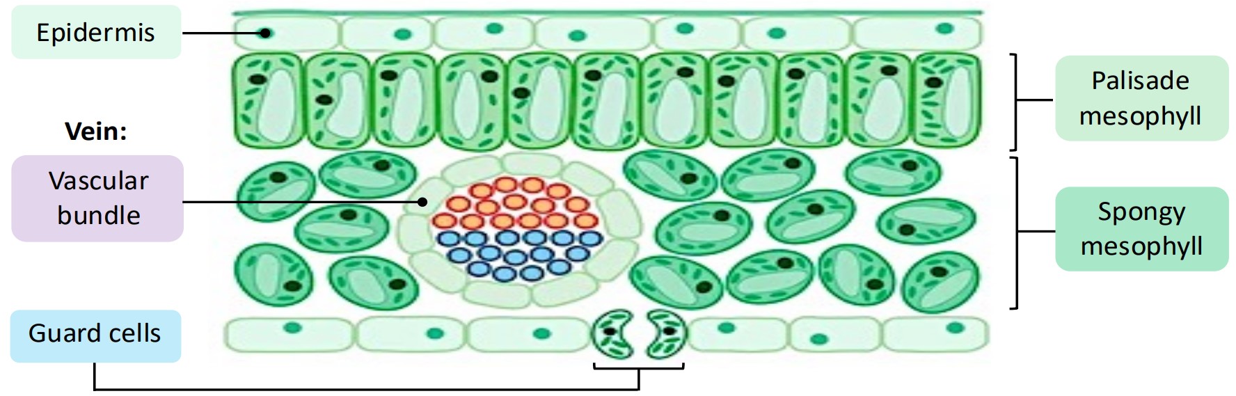
Distribution of Tissues within a Leaf (IB B3.1.8)
A leaf isn’t just a flat green sheet; it’s a highly organized structure with distinct layers, each playing a specific role:
Epidermis (Upper & Lower): A single layer of protective cells forming the leaf’s outer skin. It acts as a barrier against physical damage and pathogens.
Cuticle: A waxy, waterproof layer secreted by the epidermis, covering the outer surface. Its primary function is to prevent excessive water evaporation from the leaf surface. Imagine it as a transparent raincoat for the leaf.
Mesophyll: This is the powerhouse tissue sandwiched between the upper and lower epidermis. It’s where most photosynthesis occurs.
Palisade Mesophyll: Located just below the upper epidermis. These cells are elongated, tightly packed, and absolutely loaded with chloroplasts. Their arrangement maximizes the capture of incoming sunlight. They are the primary solar collectors.
Spongy Mesophyll: Found below the palisade layer. These cells are irregularly shaped and loosely packed, creating large interconnected air spaces. These air spaces are crucial for facilitating gas exchange (CO2 diffusion inwards, O2 diffusion outwards) between the stomata and the palisade cells.
Vascular Bundles (Veins): These run throughout the mesophyll, appearing as veins. They contain:
Xylem: Transports water and dissolved minerals from the roots up to the leaves. This water is essential for photosynthesis and replaces water lost through transpiration.
Phloem: Transports sugars (produced during photosynthesis) from the leaves to other parts of the plant where they are needed for energy or storage (like roots, fruits, or growing tips).
Diagram: Simplified Leaf Cross-Section
Example: Consider a plant growing in bright sunlight versus one growing in shade. The sun-loving plant likely has a thicker cuticle to prevent water loss and multiple layers of palisade mesophyll to maximize light absorption. In contrast, the shade plant might have a thinner cuticle and fewer palisade layers, but potentially larger leaves or more pigments to capture scarce light efficiently.
The Stem: Providing Support and Superhighways for Transport
The stem acts as the plant’s structural backbone and its critical transport network, connecting the water-absorbing roots to the photosynthesizing leaves.
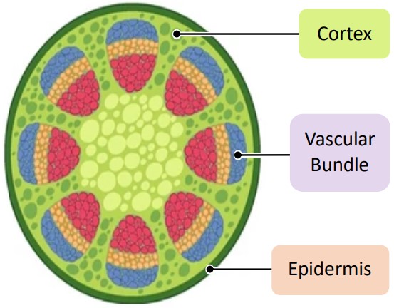
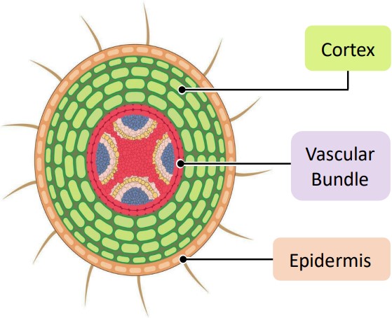
Distribution of Tissues in a Dicot Stem (IB B3.2.9)
In a transverse (cross) section, a typical dicotyledonous stem reveals an organized arrangement of tissues:
Epidermis: The outermost protective layer, similar to the leaf epidermis, often coated with a cuticle.
Cortex: Located just inside the epidermis. This layer is primarily composed of parenchyma cells and provides structural support. It can also store food reserves (like starch) and may contain chloroplasts for some photosynthesis when the stem is young and green.
Vascular Bundles: These are distinct bundles containing the transport tissues, typically arranged in a ring surrounding a central pith. This ring arrangement provides strength and flexibility.
Xylem: Positioned towards the inside of each vascular bundle. It carries water and minerals upwards.
Phloem: Positioned towards the outside of each vascular bundle. It carries sugars downwards (or upwards to developing flowers/fruits).
Cambium: A layer of actively dividing, undifferentiated cells located between the xylem and phloem in dicot stems. This meristematic tissue is responsible for secondary growth, allowing the stem to increase in diameter (get thicker) over time.
Pith: Found in the center of the stem, inside the ring of vascular bundles. It is usually composed of large, thin-walled parenchyma cells and primarily functions in food storage.
Diagram: Simplified Dicot Stem Cross-Section
Example: The sturdy trunk of an oak tree demonstrates the result of extensive secondary growth originating from the cambium within its vascular bundles. The ring arrangement provides immense strength to support the massive weight of branches and leaves, while the central pith might be relatively small compared to the extensive woody xylem developed over years.
The Root: Anchoring the Plant and Absorbing Essentials
Roots are the hidden half of the plant, anchoring it firmly in the ground while diligently absorbing water and essential mineral nutrients from the soil.
Distribution of Tissues in a Dicot Root (IB B3.2.10)
A cross-section of a dicot root shows a different tissue arrangement optimized for absorption and transport:
Epidermis: The outermost layer. A key feature here is the presence of root hairs – tiny extensions of epidermal cells that dramatically increase the surface area available for water and mineral absorption. They are like microscopic sponges reaching into the soil.
Cortex: A thick layer beneath the epidermis, primarily made of parenchyma cells. Its main role is storing food (often starch) transported down from the leaves. It also allows water absorbed by the epidermis to move towards the center.
Vascular Cylinder (Stele): The central core containing the vascular tissues, distinctly different from the stem’s ring arrangement.
Xylem: Typically forms a star-shaped (or central) structure in the very center of the root. This central placement provides tensile strength against pulling forces.
Phloem: Located in the regions between the arms of the xylem star.
Endodermis & Casparian Strip: The vascular cylinder is surrounded by a special layer called the endodermis. Embedded within the cell walls of the endodermis is the Casparian strip, a waterproof, waxy band (made of suberin). This strip blocks the passage of water between the cells (apoplast pathway), forcing water and dissolved minerals to pass through the cell membranes of the endodermal cells (symplast pathway). This ensures selective uptake – the plant controls which minerals enter the vascular system. It acts like a selective border checkpoint.
Diagram: Simplified Dicot Root Cross-Section
Example: Imagine a carrot. The orange part we eat is primarily the enlarged cortex, packed with stored sugars (food reserves). Deep inside, you’d find the central vascular cylinder, structured as described above, responsible for transporting water up to the leafy top and receiving sugars from it. The extensive root hairs (though usually lost during harvest) would have been crucial for absorbing water from the soil.
Decoding Plan Diagrams: Simplified Blueprints of Plant Tissues
Plan diagrams are simplified drawings used in biology. They show the overall layout and relative proportions of different tissues within an organ (like a leaf, stem, or root) without showing individual cells.
Purpose: To understand the basic organization and distribution of tissues quickly. They help visualize how different tissues are arranged relative to each other.
Interpretation: By looking at a plan diagram, you can infer potential functions.
A thick cortex in a root suggests significant storage capacity.
A leaf diagram with extensive air spaces in the spongy mesophyll indicates specialization for gas exchange.
The arrangement of vascular bundles (ring in stem, central in root) is clearly visible.
Key Identifier: Plan diagrams clearly show the relative positions of xylem and phloem:
Stem: Xylem inside, phloem outside within each bundle.
Root: Xylem central, phloem surrounding/between xylem arms.
Leaf (Vein): Xylem typically on the upper side (adaxial), phloem on the lower side (abaxial).
These diagrams are tools for understanding the big picture of tissue arrangement before diving into cellular details.

Conclusion: Structure Dictates Function
As we’ve seen, the arrangement of tissues in plant leaves, stems, and roots is far from random. Each structure, from the waxy cuticle to the central xylem core, is precisely adapted to perform essential functions like photosynthesis, transport, absorption, and support. Understanding this intricate relationship between structure and function is fundamental to grasping plant biology and is a cornerstone of your IB studies. Keep observing the plants around you – you now have the knowledge to appreciate their hidden complexity!
Frequently Asked Questions (FAQ)
Think of it like building blocks. Tissues are groups of similar cells working together for a specific function (e.g., xylem tissue for water transport, palisade mesophyll tissue for photosynthesis). Organs are structures made up of different tissues working together for a broader function (e.g., a leaf is an organ made of epidermal, mesophyll, and vascular tissues, all cooperating for photosynthesis and gas exchange). Roots and stems are also organs.
The arrangement reflects their primary functions. In stems, the ring arrangement of vascular bundles provides flexible strength to support the plant vertically and resist bending. In roots, the central xylem core provides strong anchorage against pulling forces from wind or gravity, while the surrounding phloem efficiently distributes sugars to the root tissues.
The Casparian strip is a waterproof barrier in the endodermis cell walls. It blocks water and dissolved minerals from flowing passively between cells into the vascular tissue. This forces water and minerals to pass through the selectively permeable cell membranes of the endodermal cells. Essentially, it gives the plant control over which minerals are absorbed into the xylem for transport to the rest of the plant, acting like a quality control checkpoint.
They specialize for different aspects of photosynthesis. Palisade mesophyll, packed with chloroplasts and near the top surface, maximizes sunlight capture. Spongy mesophyll, with its large air spaces, facilitates efficient diffusion of CO2 from the stomata to the palisade cells and allows O2 to diffuse away. This division of labor makes photosynthesis highly effective.
No, the vascular cambium responsible for significant secondary growth (increase in thickness) is characteristic of dicotyledonous plants (like trees, beans, sunflowers) and gymnosperms. Monocotyledonous plants (like grasses, corn, lilies) typically lack vascular cambium, and their vascular bundles are scattered throughout the stem, not arranged in a ring. This is why monocot stems generally don’t grow significantly thicker over time.